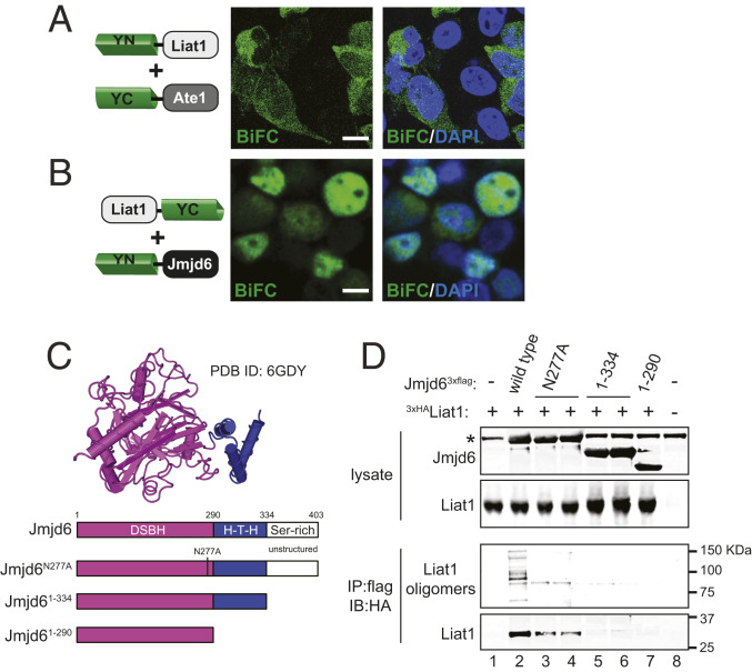
Fig. 5.
Liat1 interacts with Jmjd6. (A) BiFC between YN-Liat1 and YC-Ate1. (B) BiFC between Liat1-YC and YN-Jmjd6. Right contains DAPI. (Scale bars: 10 µm.) (C, Upper) Structure of Jmjd6 residues 1 to 334 (Protein Data Bank [PDB] ID code 6GDY). Not shown is the unstructured C-terminal serine-rich domain. (C, Lower) Schematic of wild-type Jmjd6 and its mutants used in this study. DSBH, double-stranded β-helix fold; H-T-H, helix–turn–helix. (D) Anti-flag coimmunoprecipitation of 3xHALiat1 with wild-type Jmjd63xflag or its indicated mutants. Note that higher-molecular-weight Liat1 species are detected in the presence of wild-type Liat1 but not with Jmjd6N277A despite its ability to interact with Liat1. *Antibody heavy chain. IP: immunoprecipitation using the indicated antibody. IB: immunoblot using the indicated antibody.

