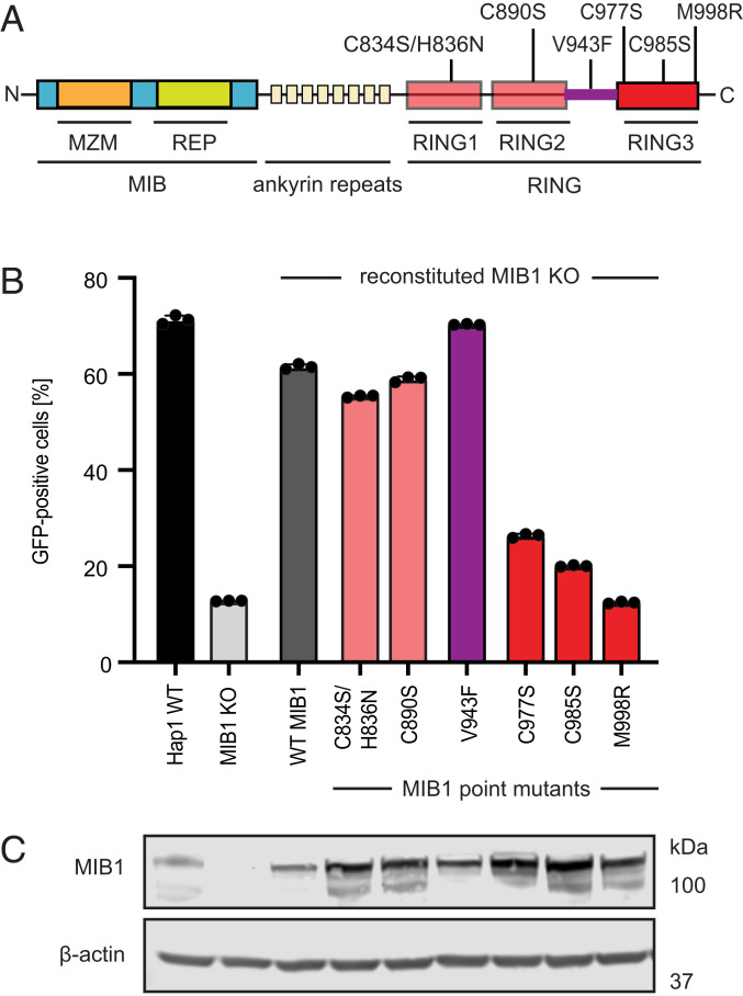
Fig. 2.
The ubiquitination activity of MIB1 is required for AdV infection. (A) Schematic of the MIB1 protein with domains and point mutations annotated. (B) AdV5rep+-GFP infection (MOI = 200 p/cell; 24 hpi) of Hap1 WT cells, MIB1 KO clone 2-1 and KO 2-1 reconstituted with panel of point mutants (annotated in A). Experiments were performed in triplicate and harvested cells were analyzed by flow cytometry and plotted as the percentage of GFP-positive cells. (C) Western blot analysis of MIB1 expression levels in Hap1 WT, MIB1 KO clone 2-1, and KO 2-1 reconstituted with MIB1 constructs in B.

