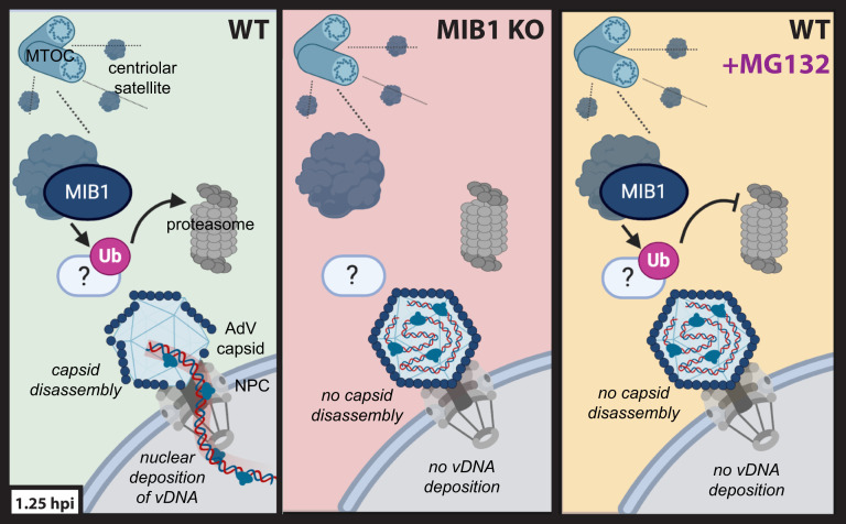
Fig. 5.
Schematic model of MIB1’s role in AdV genome deposition following capsid arrival at the nuclear pore. In WT cells (Left), MIB1, an E3 ubiquitin ligase, and centriolar satellite component, facilitates successful capsid disassembly and vDNA deposition into the nucleus through the ubiquitination and proteasomal degradation of an as-yet-unidentified target protein (white oval). In MIB1 KO cells (Middle), the MIB1 target is no longer ubiquitinated and degraded resulting in a block on capsid disassembly and genome release. Similarly, upon treatment of WT cells with MG132 (Right), the ubiquitinated MIB1 target, no longer able to be degraded by the proteasome, persists and inhibits capsid disassembly and genome release (graphic created with BioRender.com).

