Fig. 4.
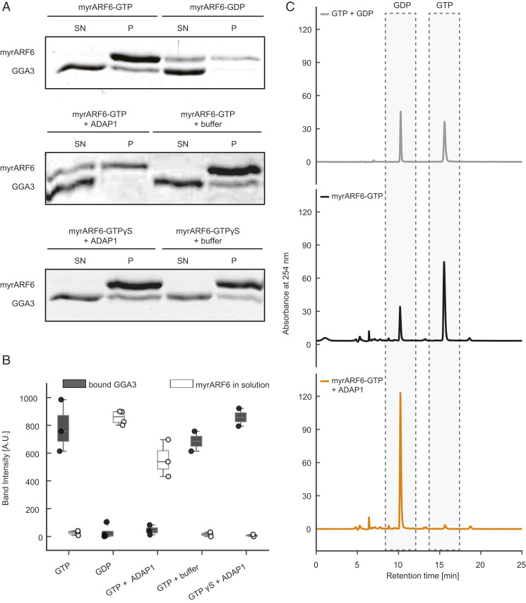
ADAP1 is an ARF6 GAP in vitro. (A, Top) To validate the assay, myrARF6 was brought into a GTP (Left) or GDP (Right) -bound state. Recruitment of GGA3 to the pellet and the amount of ARF6 in the supernatant were monitored. (A, Middle) myrARF6-GTP was incubated with 1 µM ADAP1 (Left) or ADAP1 storage buffer (Right). (A, Bottom) Instead of GTP, myrARF6 was loaded with GTPγS. (B) Quantification of ARF6 band intensity in the supernatant (white) and GGA3 band intensities in the pellet (dark gray). “Buffer” corresponds to ADAP1 storage buffer. Displayed box plots encompass the 25–75th percentiles, the midline indicates the median, the whiskers extend to show the rest of the distribution and to indicate outliers. (C) Representative HPLC chromatograms of pure GTP and GDP nucleotides (Top) compared with nucleotides extracted from PIP3 vesicle-bound myrARF6 incubated without (Middle) or with ADAP1 (Bottom).

