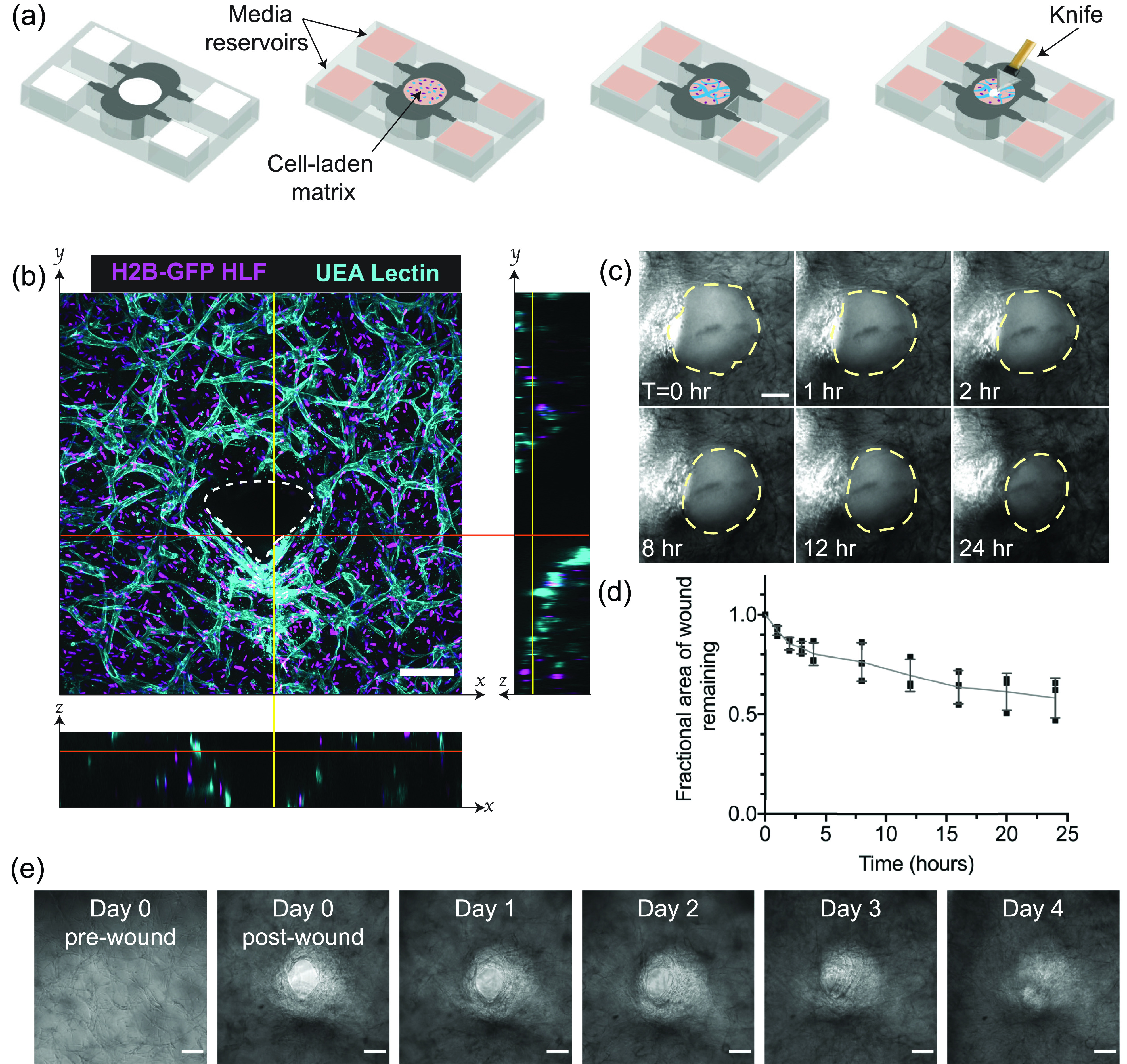
FIG. 1.

In vitro capillary beds can be injured and will heal in 3D over time. (a) Scheme for biomimetic vascularized wound healing. Endothelial cells and fibroblasts suspended within a fibrin gel are added to a PDMS mold and cultured for 3 days to develop a 3D capillary network. The devices are, then, wounded with a diamond dissection knife and imaged over time to track wound healing. (b) A wound in vascularized tissue, one day after wounding. The image is a z-projection of a 200 μm confocal stack, with cross sectional views to indicate the wound depth. White dotted lines in cross sections indicate the wound borders. The scale bar is 150 μm. (c) Brightfield image time-lapse over 24 h, with wound borders outlined with yellow dotted lines. The scale bar is 150 μm. (d) Quantification of the reduction of the wound area over the 24 h period. Error bars represent mean ± STD (n = 3). (e) Brightfield images of tissues over the course of 4 days. The scale bar is 150 μm.
