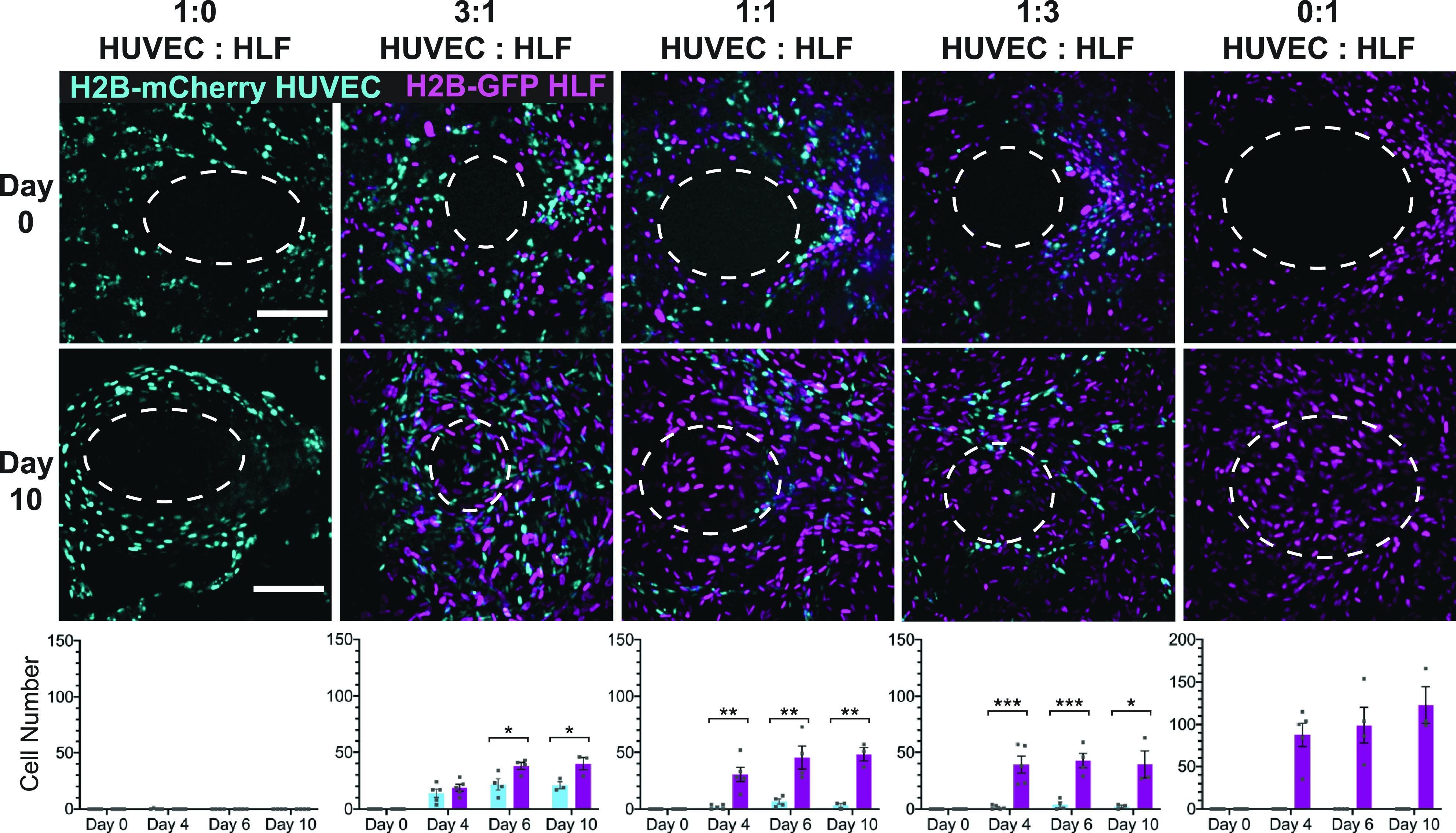
FIG. 2.

Wound closure is dependent on the presence of fibroblasts in the tissue. (Top) Projections of a confocal stack of images of the original wound area (day 0) for tissues with constant cell density but varying ratios of endothelial cells and fibroblasts. Ellipse outlines the original wound area. (Middle) Z-projections of the confocal stack of images taken after 10 days of healing, with the same ellipse overlay. (Bottom) Quantification of the number of H2B-GFP fibroblasts or H2B-mCherry HUVECs migrating into the wound over time tissues with varying starting cell ratios. The scale bar is 150 μm. Error bars represent mean ± SEM (n = 3) with 1–2 devices per condition per experiment. The significance was determined using Student's t-test; *= p < 0.05, ** = p < 0.01, and *** = p < 0.001.
