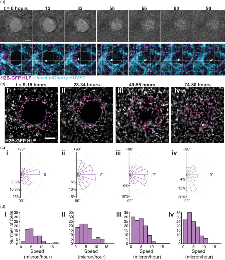
FIG. 3.
Fibroblasts migrate tangentially to the wound during contraction, while vessels fluctuate at the wound periphery. 96 h time lapse was performed to visualize healing progress over time in devices with a 1:1 ratio of HUVECs to HLFs. (a) Brightfield progression of healing over time (top). Fluorescent imaging of tissues over healing progression; the arrow indicates one vessel through the course of the time lapse (bottom). (b) Net displacement of fibroblast nuclei over 6 h segments of imaging, determined using the ImageJ Trackmate plugin. (c) Rose plots indicating the distribution of the direction of net displacement of fibroblasts for each imaging segment. (d) Histograms displaying the net velocities of individual fibroblasts over the course of each 6 h imaging segment. The scale bar is 150 μm.

