FIG. 4.

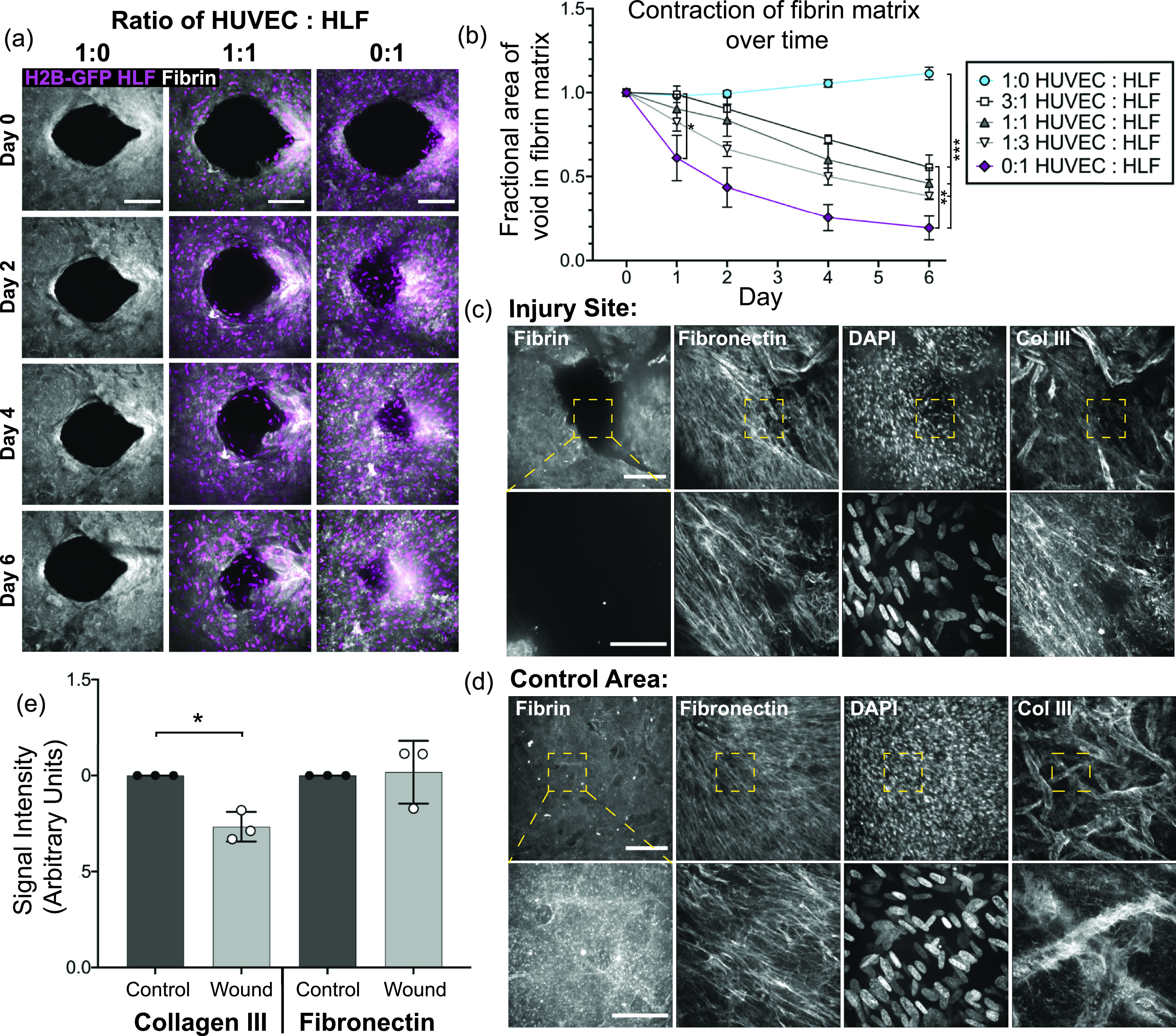
Fibroblasts remodel the original fibrin matrix during healing but deposit the provisional matrix into the wound area in order to close the gap. (a) Z-projection of the original Cy5-labeled fibrinogen matrix (gray) and HLF nuclei (magenta) over the course of healing. Tissues with HUVECs only (left), a 1:1 ratio of HUVECs to HLFs (middle), or HLFs only (right). The scale bar is 150 μm. (b) Quantification of the void area remaining in the fibrin matrix over time for devices with varying ratios of HUVECs to HLFs. Error bars represent mean ± SEM, quantified from n = 4 tissues per condition. Data were compared using ANOVA with post hoc Tukey's test; *= p < 0.05; ** = p < 0.01; and *** = p < 0.001. (c) and (d) Immunofluorescent staining of fibronectin and collagen III in tissues with the pre-labeled Cy5-fibrin matrix six days post-wounding. Images at 10× (top row; the scale bar is 150 μm) and 40× (bottom row; the scale bar is 50 μm); the area imaged at higher magnification is indicated by the yellow box. (c) Images taken directly at the site of injury and (d) at a control site away from the wounded area. (e) Quantification of the fibronectin and collagen III staining intensity within the healed region or within the control tissue, quantified from 5 to 6 regions of interest in three different tissues from the 10× images as shown in (c) and (d). Data were compared with a paired t-test, * = p < 0.05.
