Fig. 3.
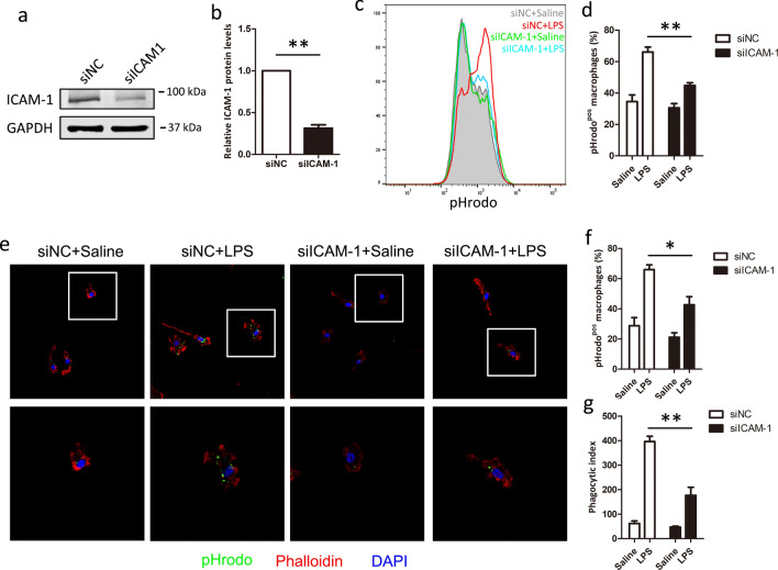
Silencing of ICAM-1 expression reduces LPS-induced phagocytosis in macrophages. BMDM cells were transfected with si-NC or si-ICAM-1 for 48 h. a, b The efficiency of silencing was evaluated by western blot after 48 h. GAPDH was used as quantitative controls. BMDM cells transfected with si-NC or si-ICAM-1 were treated with LPS for 24 h to assay the macrophage phagocytosis. c, d Macrophage phagocytosis was analyzed using pHrodo by flow cytometry. c Representative histogram illustrating the detection of pHrodo associated macrophages. d Percentage of pHrodo-positive (pHrodopos) macrophages is shown. e–g Macrophage phagocytosis was analyzed using pHrodo by confocal macroscopy. e Representative image of macrophages phagocytosis. Percentage of phagocytosis (f) and Phagocytic index levels (g) are shown. Each bar represents the mean ± SD based on three independent experiments. *P < 0.05, **P < 0.01

