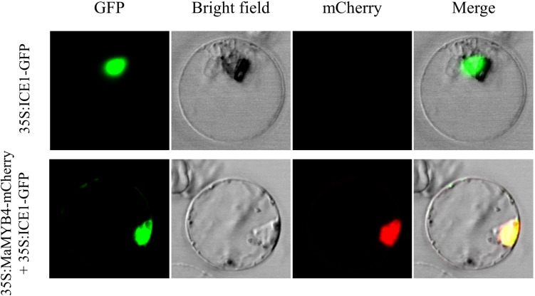
FIGURE 2.
Subcellular localization analysis of MaMYB4. The open reading frames of MaMYB4 and ICE1 (used as the nucleus marker) were in framed with GFP and mCherry N-terminus, respectively. The obtained constructs driven by 35S promoter were transient expressed in rice protoplasts and visualized by fluorescence microscopy. The GFP fluorescence signals indicated the localization of nucleus. Overlay images show colocalization of GFP and mCherry signals.

