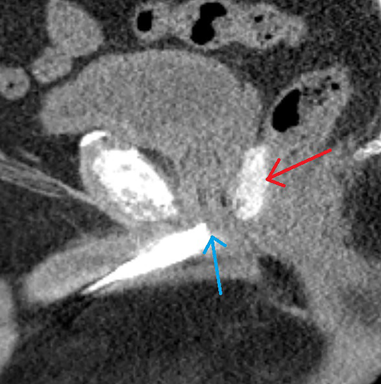
Figure 2.
A 69-year-old male with intermediate risk prostate cancer had an implantable cardiac pacemaker which precluded magnetic resonance imaging for treatment planning. Thus he underwent placement of SpaceOAR Vue™ iodinated rectal spacer and a urethrogram CT image was obtained for treatment planning: Treatment planning sagittal computed tomography urethrogram images demonstrate the radiopaque spacer between the prostate and rectum (red arrow), as well as the beak of the urethrogram (blue arrow).

