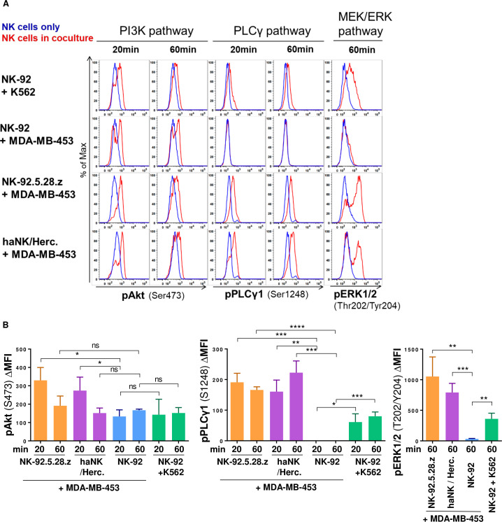
Figure 7.
Resistant MDA-MB-453 cells induce PI3K signaling in NK-92 but fail to trigger PLCγ or MEK/ERK activation, which is rescued by car or ADCC. NK cells were mixed with cancer cells for the indicated time periods or kept alone. Phosphorylation of the indicated signaling molecules in NK cells was determined by flow cytometry. NK cells were discriminated by CD56 staining. PI3K, PLCγ and MEK/ERK pathways were analyzed by assessing pAkt (Ser473), pPLCγ1 (Ser1248) and pERK1/2 (Thr202/Tyr204), respectively. (A) Histograms show in blue NK cells only, and in red NK cells cocultured with the indicated target cells. (B) Mean fluorescence intensity (MFI) data from 3 independent experiments. Mean values ±SD are shown. ERK, extracellular signal-regulated kinase; MEK, mitogen-activated protein kinase; NK, natural killer; PI3K, phosphoinositide 3-kinase; PLCγ, phospholipase C-γ. ns, not significant; *P<0.05, **P<0.01, ***P<0.001, ****P<0.0001.

