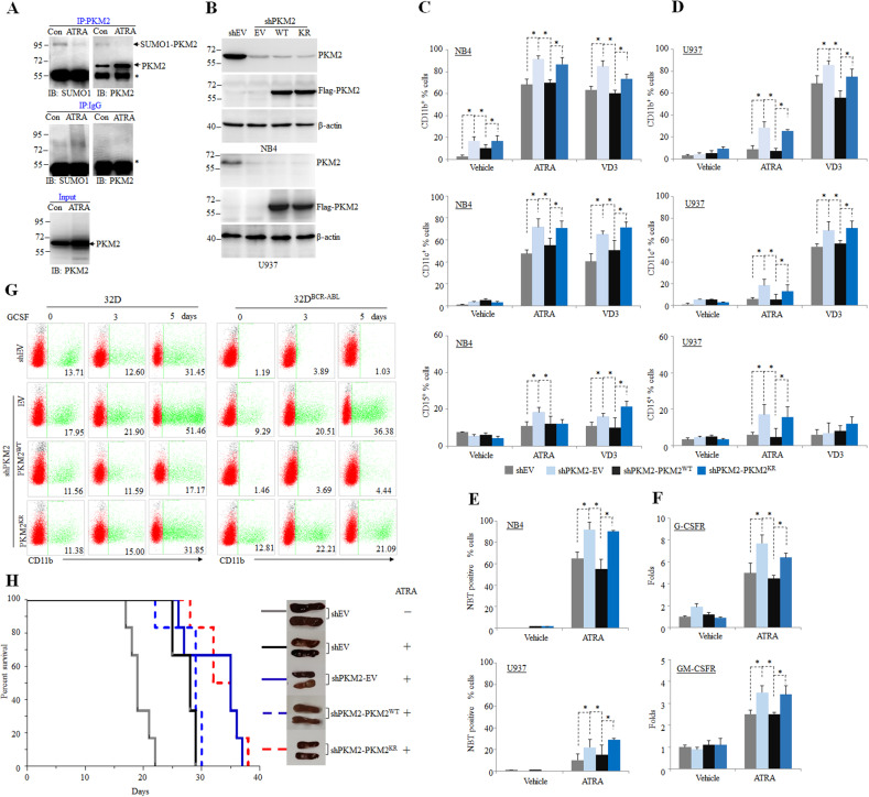
Fig. 4. SUMOylation of PKM2 inhibits myeloid differentiation.
A NB4 cells were treated with 10−8 M ATRA for 3 days followed by IP with anti-PKM2 antibody. The SUMO1 modification were analyzed by western blot using anti-PKM2 antibody. B Western blot analysis of NB4 and U937 cells in which endogenous PKM2 was stably knocked out and WT PKM2 (WT) or K270R mutant (KR) were re-expressed. C–E The NB4 and U937 rescue cell lines were treated with 10−8 M ATRA or 100 nM VD3 for 3 days and the CD11b-positive, CD11c-positive, or CD15-positive cells were measured by flow cytometry (C, D) and NBT positive cells were counted (E). *p < 0.05 between the line-pointed group. F The mRNA level of GCSFR or GMCSFR quantified by real-time RT-PCR were shown. G The 32D or 32DBCR-ABL rescue cell lines were treated with 50 ng/ml GCSF for indicated times and the CD11b-positive cells were counted by flow cytometry. Data are shown as the mean ± SEM of three independent experiments. H Human NB4 cells (1 × 10⁷) were injected via tail vein into sub-lethally irradiated NSG mice followed by treatment with vehicle or ATRA (10 mg per kg body weight, intraperitoneally) daily for five continuous days a week. The survival data were collected. The representative pictures of spleens from each group were shown on the right.

