Fig. 6.
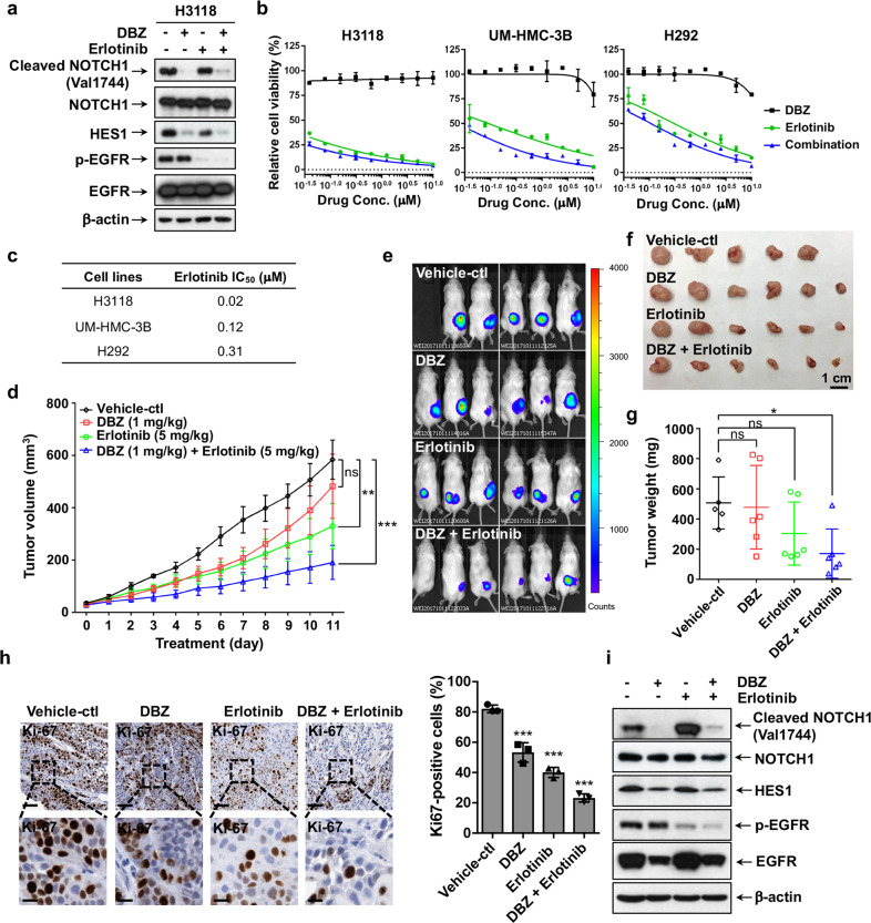
Co-inhibition of Notch and EGFR signaling synergistically inhibited the growth of MEC xenografts. a H3118 cells were treated with either DBZ (1 μM), Erlotinib (1 μM), or in combination for 24 h, and the treated cells were analyzed for Notch and EGFR signaling status by Western blot analysis using the antibodies as indicated. b Cell viability assays were performed on human MEC cells treated with various concentrations of DBZ, Erlotinib or in combination for 72 h. c The IC50 values of Erlotinib were shown. The IC50 of DBZ was not applicable as no inhibitory effect was observed. d–g NOD.SCID mice were subcutaneously inoculated with luciferase-expressing H3118 MEC cells. When the xenografts grew to ~50 mm3, mice were treated with vehicle control (n = 5), 1 mg/kg DBZ (n = 6), 5 mg/kg Erlotinib (n = 6), or 1 mg/kg DBZ plus 5 mg/kg Erlotinib (n = 6) via IP injection (DBZ) or oral gavage (Erlotinib) daily for 11 days. The tumor volumes were measured daily after treatment (d). Bioluminescent imaging of the xenograft tumors (e), tumor size (f), and tumor weight (g) were presented on the final day of treatment. h Representatives of Ki-67 staining of H3118-luc xenografts from each treatment group were shown (upper bar = 100 μm, lower bar = 25 μm). ImageJ was used to calculate Ki-67-positive cells in IHC staining results and sections of individual tumors (n = 3) in each group were analyzed. i Western blotting was performed to determine the levels of cleaved and total NOTCH1, HES1 as well as the phosphorylated and total EGFR post-drug treatments. The mice were given a boost administration of DBZ (5 mg/kg) and Erlotinib (25 mg/kg) either alone or in combination 2 h prior to euthanasia (*p < 0.05; **p < 0.01; ***p < 0.001)

