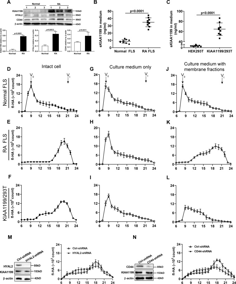
Fig. 3. Degradation of exogenous fl-HA by intact cells and their media without or with the addition of cell membrane fractions.
A Western blot analysis of KIAA1199, HYAL2, and CD44 protein in FLS from normal subjects (n = 3) and RA patients (n = 3). The relative expression of protein was calculated by the gray ratio of protein band to β-actin. B, C Comparison of sKIAA1199 in the culture media of normal FLS and RA FLS (B), HEK293T and KIAA1199/293T cells (C). Normal FLS and RA FLS were isolated from synovial tissues of normal subjects (n = 3) and active RA patients (n = 3), respectively. D–L The HA-degrading activity of intact normal FLS, RA FLS, and KIAA1199/293T cells (D–F), their culture media without (G–I) or with cell membrane fractions (J–L). Arrow head indicated the positions of the void volume (Vo) and the total volume (Vt) of the column. M, N The effect of HYAL2 knockdown (M) or CD44 knockdown (N) in RA FLS on the HA-degrading activity of membrane-bound sKIAA1199. All experiments were performed at least in triplicates, the data were presented as mean ± SD.

