Figure 1.
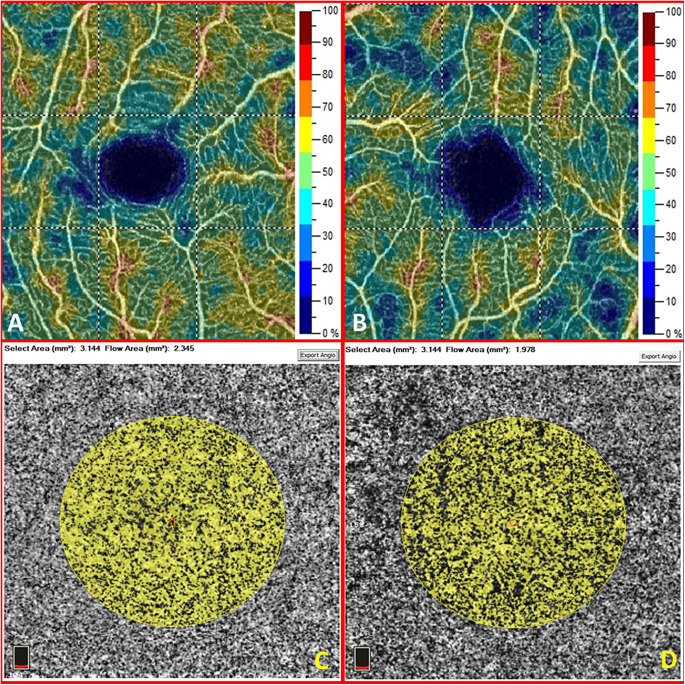
The macular superficial capillary plexus vessel density map (A) and the macular choriocapillaris vascular perfusion area map (C) of the right eye in a healthy 66-year-old woman. The macular superficial capillary plexus vessel density map (B) and the macular choriocapillaris vascular perfusion area map (D) of the left eye in a 55-year-old woman with glaucoma. The corresponding whole image vessel density and retinal thickness are significantly reduced in eyes with glaucoma (49.3 vs. 44.1% and 326 vs. 287 microns in this case). In the selected area of 3.144 mm2 centered in the fovea, the choriocapillaris perfusion area is reduced in this case (2.345 vs. 1.978 mm2). This difference, however, is not statistically significant when considering all glaucomatous eyes of the study versus the others.

