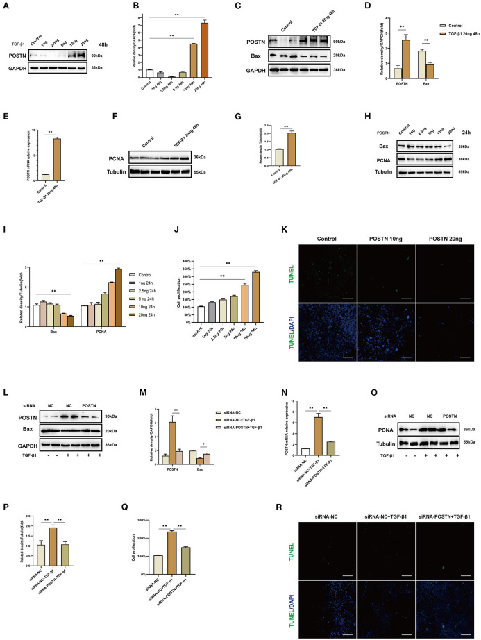
Figure 7.
High levels of POSTN enhance the proliferation and suppress apoptosis in MMCs. (A,B) Immunoblot analysis and quantification of POSTN in MMCs treated with indicated concentrations of TGF-β1 for 48 h. (C–I) Immunoblot analysis and quantification of POSTN, BAX, and PCNA in MMCs treated with 20 ng/ml TGF-β1. (H,I) Immunoblot analysis and quantification of BAX and PCNA in MMCs treated with indicated concentrations of recombinant POSTN protein for 24 h. (J) Proliferation of MMCs as detected with the CCK-8 assay. (K) Detection of apoptotic cells with the TUNEL assay. Scale bar: 50 μm. (L,M) siRNA-mediated POSTN knockdown in MMCs. Immunoblot analysis and quantification of POSTN and BAX levels in TGF-β1–treated MMCs after POSTN knockdown. (N) POSTN mRNA level in TGF-β1–treated MMCs after POSTN knockdown. (O,P) Immunoblot analysis and quantification of PCNA in TGF-β1–treated MMCs after POSTN knockdown. (Q,R) Detection of proliferation and apoptosis with the CCK-8 and TUNEL assays, respectively, in TGF-β1–treated MMCs after POSTN knockdown. Scale bar: 50 μm. Data are presented as mean ± SEM (n = 3). *P < 0.05, **P < 0.01.

