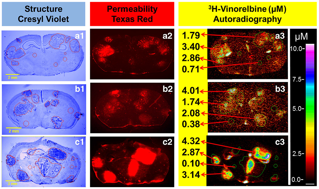
Fig. 3.

Experimental brain metastases exhibit heterogeneous permeability and uptake of vinorelbine. Representative coronal brain section images at 0.5–8 h following i.v. administration of 3H-vinorelbine. (a1-c1) MDA-MB231BR brain metastasis localization and identification based on cresyl violet staining, yellow scale bar represents 2 mm. (a2-c2) Texas red fluorescence shows heterogeneous distribution and was used as marker of passive permeability in uninvolved brain and metastases. (a3-c3) Heterogeneous distribution of 3H-vinorelbine in brain metastases by QAR. Images across a row are of the same brain section.
