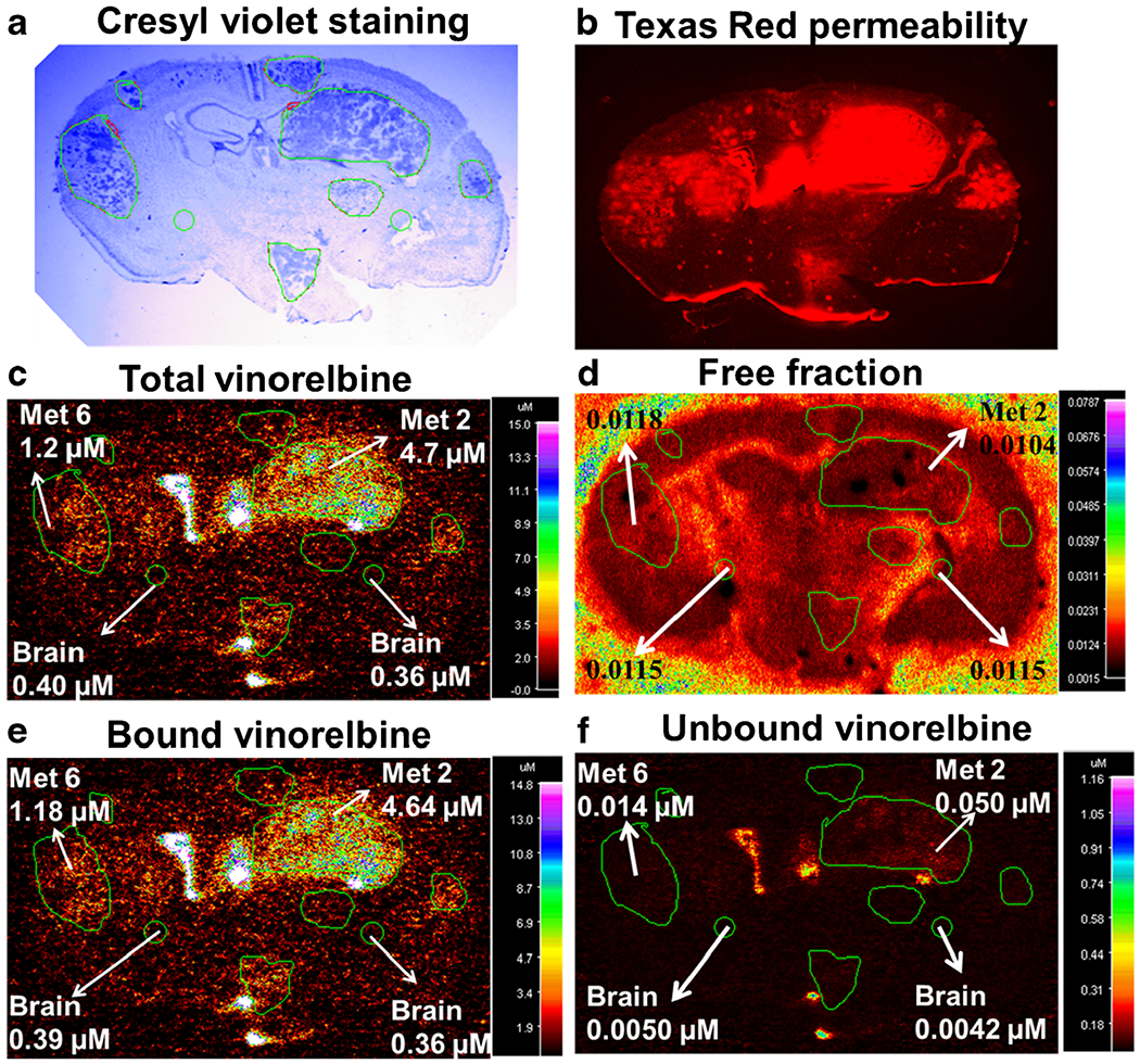
Fig. 7.

Vinorelbine total, bound and unbound concentration in MDA-MB-231BR experimental brain metastases of breast cancer. Representative images for determination of unbound vinorelbine concentration in 20 μm coronal brain sections from mice harbouring metastases 2 h after injection of vinorelbine. a Cresyl violet staining of brain section for tumor localization. b Fluorescence image of Texas red permeability. c QAR image of in vivo total vinorelbine. d QAR image of in vitro free fraction. e Bound vinorelbine concentration. f Unbound vinorelbine concentration. Images a-c and e-f are of the same brain section, while d is from the adjacent bran section. Images d-f were obtained following the procedure illustrated in Fig. 1 using MCID software.
