
Figure 3.
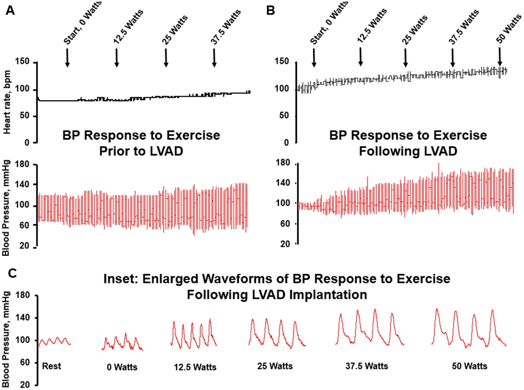
Example tracing of the heart rate and blood pressure response to exercise in a 46yo man with history of advanced HFrEF. (A): exercise prior to LVAD implantation; (B): exercise test following LVAD implantation; (C): inset demonstrates enlarged BP waveforms from B, emphasizing the early increase in pulsatility following initiation of exercise. Note dicrotic notch in arterial waveform, demonstrating aortic valve opening.