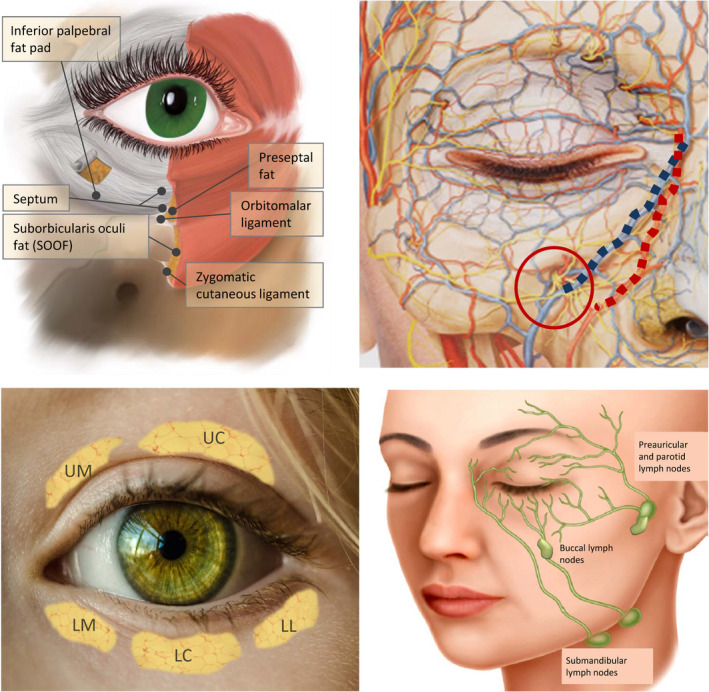
Figure 1.

Anatomical structures involved in the formation of the tear trough deformity. This is located between the palpebral and orbital areas of the orbicularis oculi muscle, and the location of the nasojugal fold corresponds to the lower boundary of that muscle. Top left: Anatomy of the periocular area. Top right: Image of the vascular and nerve structures of the area. The blue line highlights the angular vein and the red the angular artery; the red circle indicates the emergence of the infraorbital nerve. Bottom left: Location of the fat pads: upper medial (UM), upper central (UC), lower medial (LM), lower central (LC), and lower lateral (LL). Bottom right: lymph drainage of the periocular area
