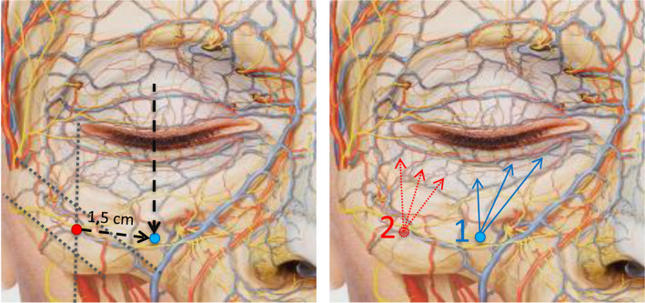
Figure 6.

Left. Diagram locating the entry point for needle injection. Left. The initial reference point (red) is located using the references described for the cannula injection in Figure 4. Starting from it, the needle insertion point (blue) will be located by advancing toward the projection of the midpupillary line, at approximately 1.5 cm, tracing a downward diagonal parallel to the orbital rim. It is important not to advance more toward the vascular/nerve bundle. Right. The needle is inserted at point 1, and the product is deposited according to the linear retrograde fanning technique, starting with the points indicated by the blue arrows. If the patient also needs treatment of the palpebromalar groove, the needle will be inserted at point 2, injecting using the same technique (dotted red arrows)
