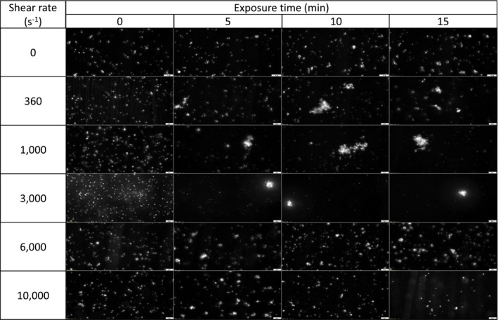
TABLE 2.
Captured images (× 40 objective) of platelet aggregates by microscopy at different shear rates (0‐10,000 s−1) and exposure time (0‐15 min). Platelet aggregates were not observed in no‐shear environments (<50 μm2). During low shear conditions ranging from 360 to 3,000 s−1, large platelet aggregates were observed (> 200 μm2); whereas smaller platelet aggregates (< 100 μm2) were observed in high shear conditions from 6,000 to 10,000 s−1