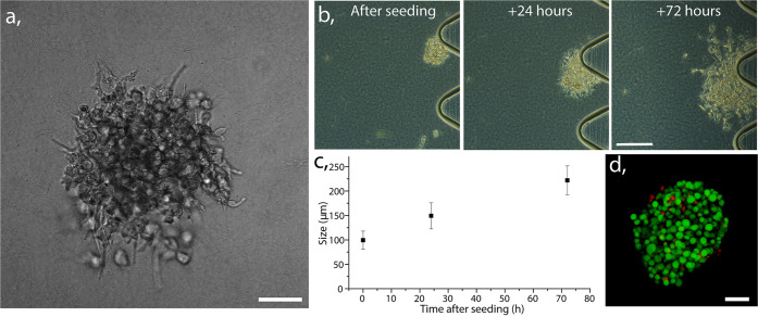
Figure 3.
(a) Transmission image of collagen gel-embedded HeLa cells conformed into 3D spheroid. Scale bar 50 μm. (b) Transmission images demonstrating proliferation of a gel-embedded spheroid from the preparation up to 3 days in perfused culture. Scale bar 100 μm. (c) Graph demonstrating size progression of gel embedded spheroids after 1 and 3 days of perfused culture, calculated based on diameter measured in 10 different chips, average with standard error bars. (d) Confocal image of stained HeLa spheroid with calcein (live cells: green) and Propidium iodide (dead cells: red). Scale bar 50 μm.

