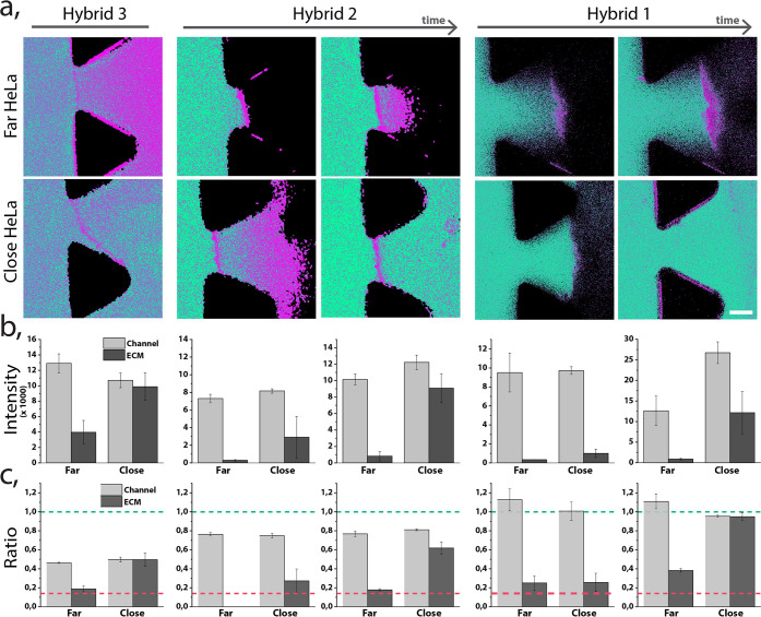
Figure 6.
Stability of hybrids 1–3, perfused through a tumor blood vessel models with different EB distance to the HeLa spheroid (close: < 400 μm, far: > 1 mm). (a) Ratiometric confocal images of the different hybrids perfused through the chip in two regions. Hybrids 1 and 2 are shown at two different time points: less than 15 min and after 30 min of continuous perfusion; hybrid 3 demonstrated at one time point due to its rapid penetration through the EB. Scale bar 75 μm. (b) Summed up fluorescence intensity originating from the monomer and the micelle channels. Intensity was measured in the blood vessel model channel and in the ECM region. (c) Normalized ratio of fluorescence signal between micellar and monomer form for each hybrid and in each region. Green dashed line indicates the ratio of fully formed micelles in equilibrium, and the magenta dashed line indicates the ratio of fully disassembled (monomer) form.

