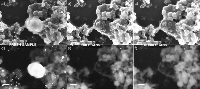
Figure 10.
IL-SEM imaging of the Au/C sample during AST in 0.05 M H2SO4 + 10–2 M Cl–: (a,d) fresh sample; (b,e) after 500 scans; (c,f) after 10,000 scans. Scale bars correspond to 100 nm. Upper row of images was obtained using the in-lens detector, while the lower row was obtained using the SE2 detector.

