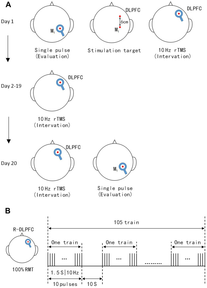
Figure 1.
TMS protocol for patients. (A) Single pulse TMS-MEP recordings before the protocol were used to collect data and locate the M1. DLPFC was 6 cm anterior to the M1 paralleled to the sagittal line. TMS-MEP evaluations were not conducted on days 2-19. Single pulse TMS-MEP recorded immediately after the 20 sessions were used to assess protocols. (B) Illustration of a single daily session of stimulation consisted of 10 Hz trains for 1.5 sec. These were repeated 105 times with an inter-train interval of 10 sec. Total sessions were 20 min and 8 sec. TMS-MEP, transcranial magnetic stimulation-motor evoked potentials; DLPFC, dorsolateral prefrontal cortex.

