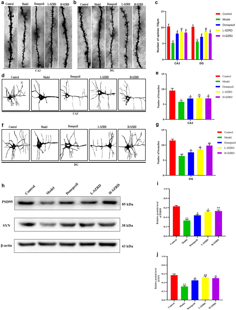
Fig. 3.
Effect of SZRD on the synaptic plasticity of APP/PS1 transgenic mice. a Representative Golgi-Cox staining in the hippocampus of CA3 after treatment with and without SRZD for 4 weeks (magnification, × 400; scale bar represents 20 µm). b Representative Golgi-Cox staining in the hippocampus of DG after treatment with and without SRZD for 4 weeks (magnification, × 400; scale bar represents 20 µm). c The number of spines in the areas of CA3 and DG. d Representative Golgi-Cox staining in the hippocampus of CA3 after treatment with and without SRZD for 4 weeks (magnification, × 400; scale bar represents 20 µm). e The number of spines in the areas of CA3. f Representative Golgi-Cox staining in the hippocampus of DG after treatment with and without SRZD for 4 weeks (magnification, × 400; scale bar represents 20 µm). g The number of spines in the areas of DG. h Examples of original Western blotting bands showing expressions of PSD95 and SYN in the hippocampus. i Relative protein level of PSD95. j Relative protein level of SYN. The data are presented as mean ± SEM (n = 4 in each group). **P < 0.01 vs. Control group; #P < 0.05 and ##P < 0.01 vs. Model group

