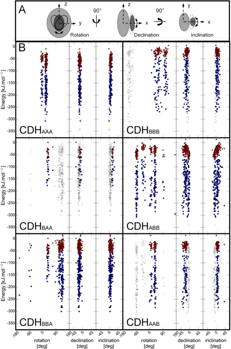
Figure 5.
Orientation of CYT to DH in docking poses. (A) Schematic representation of evaluated angles. (B) From a total of 200 docking poses for each CYT–linker–DH pair the angle of rotation, declination, and inclination were measured in regard to its deviation from the crystal structure of the closed-state conformation of M. thermophilum CDH (PDB ID: 4QI6). The electrostatic (red) and van der Waals (blue) binding energies for each pose are given in kJ mol–1. Docking poses in wild-type CDHAAA and wild-type CDHBBB are compared to docking poses of chimeric CDHBAA, CDHABB, CDHBBA, and CDHAAB.

