Fig. 2.
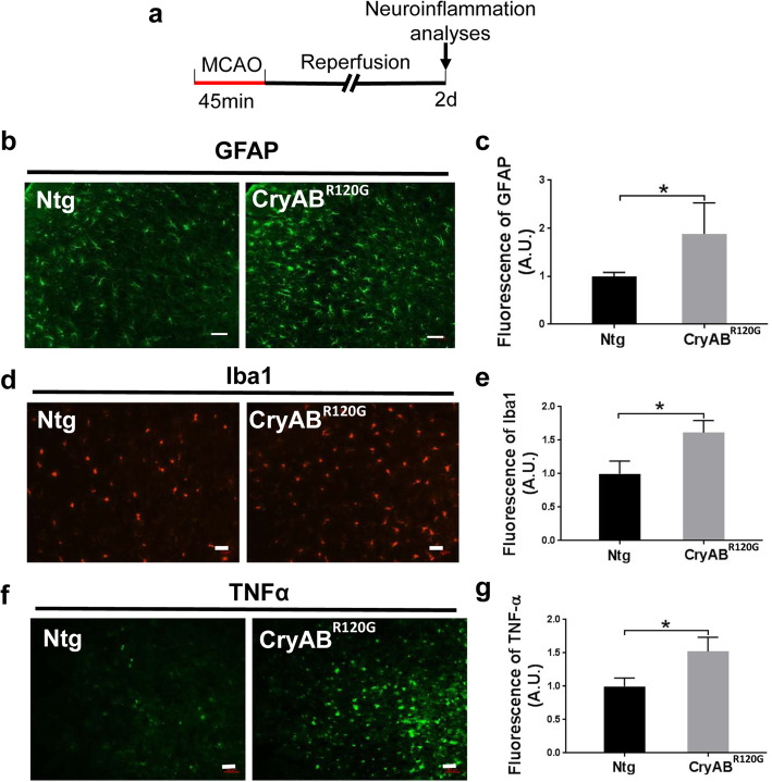
Cardiomyocyte-restricted expression of misfolded CryABR120G protein enhanced I/R-induced neuroinflammation in the brain. a Diagram of the experimental design. b Immunofluorescence staining of GFAP in the peri-infarct zone brain of mice after MCAO. Scale bar, 50 μm. c Quantitative analysis of (b). d Immunofluorescence staining of Iba1 in the brain of mice after MCAO. Scale bar, 50 μm. e Quantitative analysis of (d). f Immunofluorescence of TNFα in the cortical infarct brain of mice after MCAO. Scale bar, 50 μm. g Quantitative analysis of (f). Data are shown as mean ± SD; for immunostaining, 10–16 sections per animal were imaged and analyzed using the ImageJ software. N = 3–4. *p < 0.05, **p < 0.01

