
Figure 5.
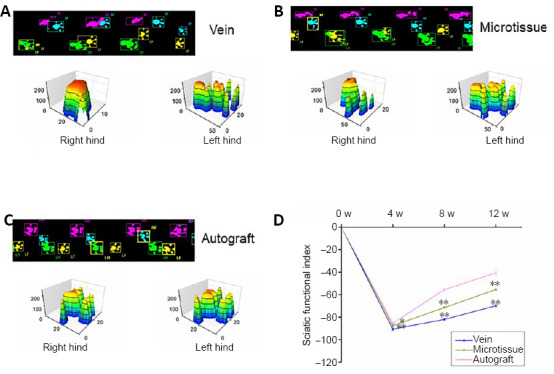
Recovery of sciatic nerve function in rats given vein/nerve microtissue hybrid scaffold graft.
(A–C) Real-time movie images and 3D footprints in the vein, microtissue, and autograft groups at 12 weeks after transplantation. In the 2D figures, blue represents the right forelimb, yellow represents the left forelimb, pink represents the right hind limb (operative side), and green represents the left hind limb (normal side). The 3D footprints show that the contact area between the toe and the ground on the operative side (right side) was decreased in each group compared with the normal side (left side). However, the length and width of the toe prints on the operative side in the microtissue group were improved compared with the vein group and similar to the autograft group. (D) The static sciatic index values at different time points after surgery (n = 8 for each group per time point). Data are expressed as the mean ± SD. **P < 0.01 (one-way analysis of variance followed by Tukey’s post hoc test).