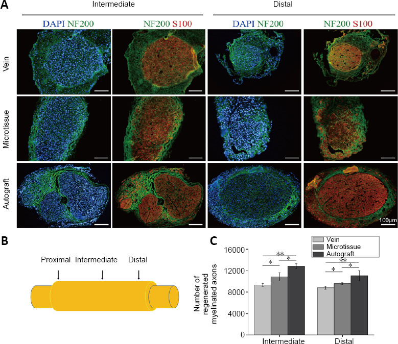
Figure 7.

Immunofluorescence staining of nerve grafts in rats with vein/nerve microtissue hybrid scaffold transplant at 12 weeks post-surgery.
(A) Labeling of histological cross-sections for S100 (red, Alexa Fluor-594), NF200 (green, Alexa Fluor-488) and DAPI (blue) in rats implanted with each nerve guide conduit. Scale bars: 100 μm. (B) Schematic diagram of the nerve graft. (C) Total number of regenerated myelinated axons in the intermediate and distal region of a graft in the three groups at 12 weeks post-surgery (n = 8 randomly selected fields for each group). Data are expressed as the mean ± SD. *P < 0.05, **P < 0.01 (one-way analysis of variance followed by Tukey’s post hoc test). DAPI: 4′,6-Diamidino-2-phenylindole; NF200: neurofilament 200.
