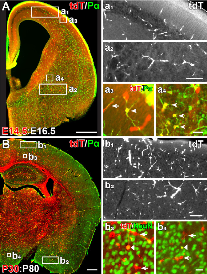
Figure 1.

NG2 glia in embryonic and adult brain.
The distribution of embryonic and adult NG2 glia in the brain demonstrated in NG2-CreERT2Huang KIxRosa26-tdTomato mice. In the embryonic brain, of the NG2 gene started in a small portion of OPCs in the ventral brain. When Cre activity was induced at E14.5 and analyzed at E16.5 (E14.5:E16.5), tdTomato (tdT) expression was found in PDGFRα+ OPCs (therefore termed NG2 glia) and vascular pericytes (A, a1–a4). Most tdT+ NG2 glia were detected in the ventral brain (A, a2, and a4, arrowheads) and few were found in the dorsal cortex (A, a1, and a3, arrowheads). Nevertheless, vascular tdT+ pericytes were observed in the whole brain (A, a1–a4, arrows). In adult brain at P30:P80, tdT+ NG2 glia were found equally distributed in the brain, irrespective of dorsal or ventral regions (B, b1, b2). Some NeuN+ tdT+ cells with typical neuronal morphology were sporadically located in the cortex (b3, triangle). However, only few tdT+ cells (~0.5%, 5 out of 946 cells analyzed from three mice) were NeuN immunopositive in the hypothalamus (b4, triangle). Scale bars: 500 μm in A and B; 100 μm in a2, and b2; 20 μm in a4, and b4 (unpublished data from Huang et al., 2014, 2019).
