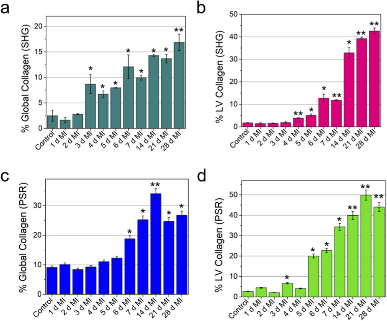
Fig. 3.
Global (whole heart) and local (LV region) quantification of collagen deposition showing the progression of fibrosis after MI. Quantification of collagen deposition using SHG images (a) calculated from whole heart, (b) calculated from the LV region. Quantification of collagen deposition using picrosirius red staining (c) calculated from whole heart, (d) calculated from the LV region. The collagen percentage was calculated as the ratio of the area occupied by the collagen to the total area of the tissue (a, c) or the area occupied by the collagen to the area of the LV region (b, d). For each time point the collagen percentage were averaged over two separate heart sections. Heart from separate mice were used for each time point. SHG and PSR imaging were performed on heart tissue collected from separate mice. *p < 0.05, **p < 0.001, one-way ANOVA.

