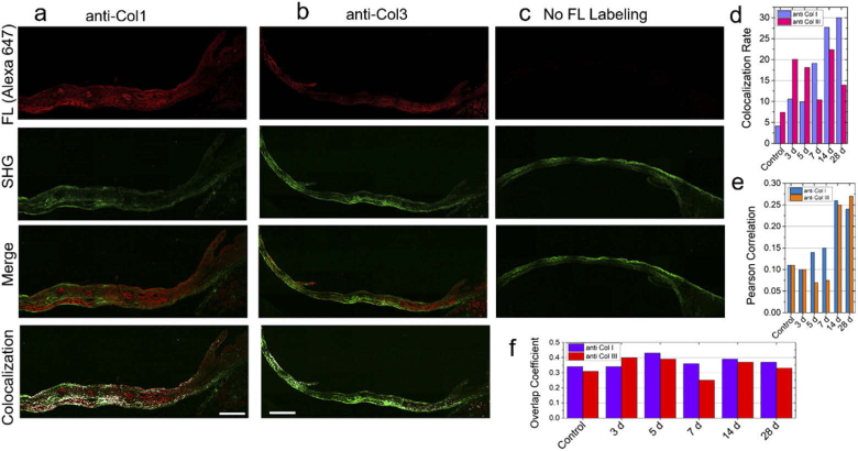
Fig. 4.
Comparison of immunohistochemistry (IHC) and SHG imaging after MI. Representative image of Alexa647-labeled (a) collagen type I (anti-col1), (b) collagen type III antibodies (anti-col3), and (c) no fluorescence in heart tissue section post 4 week (28 d) of MI injury. The SHG images of the same tissue immunolabeled with anti-col1 and anti-col3 are shown in the second panel under a-c captured employing 25x, 0.95 NA water objective lens. The overlap images of the fluorescence and SHG are shown in the third panel under a-c. The colocalization analysis of the SHG and fluorescence images are shown in the fourth panel. (d) Colocalization rate, (e) Pearson correlation coefficient, and (f) Overlap coefficient comparing anti-col1 and anti-col3 at different time points after MI. Scale bar: 250 µm.

