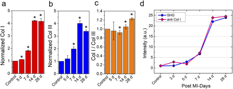
Fig. 5.
Time-dependent distribution of different isoforms of myocardial collagen after MI. (a) Normalized collagen type I expression with respect to uninjured hearts at different time points after MI. (b) Normalized collagen type III contents with respect to uninjured heart at different time points post-MI. (c) ratio of Col I to Col III distribution during the course of progression of fibrosis monitored over 4 weeks’ time period. (d) Comparison of variation of SHG and fluorescence signal as a function of days after MI showing a good correlation between SHG and immunohistochemistry studies. For each time period, two separate heart sections were analyzed. *p < 0.05, one-way ANOVA.

