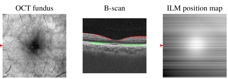
Fig. 6.
Layer segmentation in a representative X-fast scan from a 28 y/o healthy subject. From left to right: OCT fundus image, B-scan at the position of the arrow in the fundus image, with segmented ILM and EZ / IS-OS, ILM position map showing deeper ILM positions in white. The image is consistent with the expected foveal contour.

