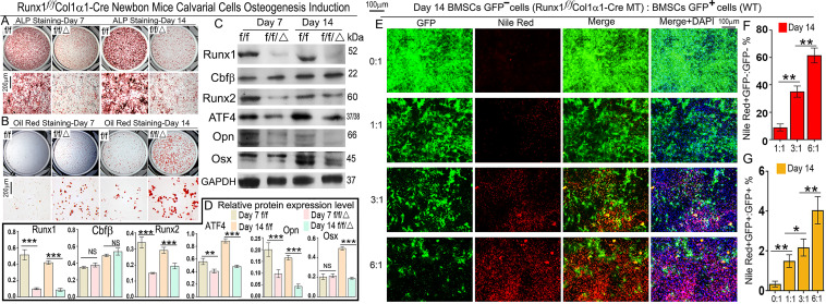
Fig 3. Runx1 deficiency in primary calvarial cells cultured from Runx1f/fCol1α1-Cre mice inhibits osteoblastogenesis and promotes adipogenesis.
(A, B) Calvarial cells from Runx1f/fCol1α1-Cre (ff/Δ) and control (ff) newborn mice were applied to osteoblastogenesis assays (A) ALP and (B) Oil red staining. (C) Protein levels of Runx1, Cbfβ, Runx2, Atf4, Opn, and Osx were analyzed by Western blot analysis. (D) Quantification of western blot results in C. (E) Runx1f/f Col1α1-Cre;GFP- and GFP+ bone marrow MSCs were mixed together in different ratios and cultured in osteogenic medium. Adipocytes were labeled by Nile Red and couterstained by DAPI on Day 14. (F, G) Quantification of (F) Nile Red+GFP-/GFP- and (G) Nile Red+GFP+/GFP+ ratios in E. Results are expressed as mean ± SD, n ≧ 3 in each group. *p < 0.05, **p < 0.01, ***p < 0.001.

