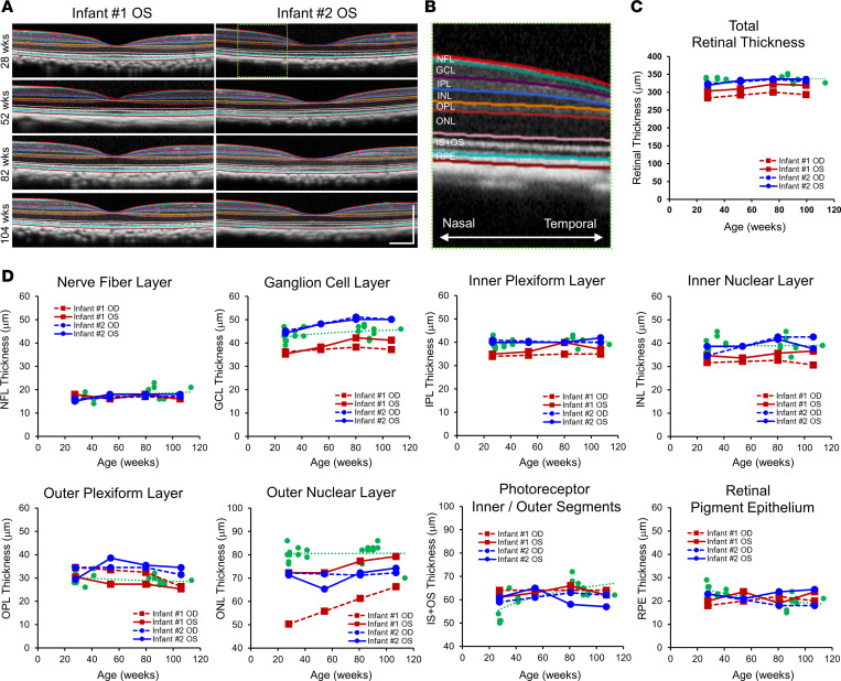
Figure 5. Retinal thinning in ZIKV-infected infant macaques.
(A) Representative SD-OCT images of 2 infant macaques (no. 1 and no. 2) exposed to ZIKV infection in utero, with semiautomated segmentation of retinal layers. (B) Magnified view of the green dashed region of the SD-OCT image in A showing the retinal layers of the nasal parafoveal area, including the NFL, GCL, IPL, INL, OPL, ONL, IS+OS, and RPE. (C) Total retinal thickness and (D) individual retinal layer thicknesses measured from right (OD, dashed lines) and left (OS, solid lines) eyes from ZIKV-infected infant no. 1 (red) and no. 2 (blue), compared with eyes from healthy animals (green) and their trendline across similar ages. ZIKV, Zika virus; SD-OCT, spectral domain–optical coherence tomography; NFL, nerve fiber layer; GCL, ganglion cell layer; IPL, inner plexiform layer; INL, inner nuclear layer; OPL, outer plexiform layer; ONL, outer nuclear layer; IS+OS, photoreceptor inner and outer segments; RPE, retinal pigment epithelium; OD, right eye; OS, left eye.

