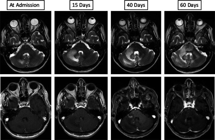
Fig. 1.
Cerebral magnetic resonance imaging (cMRI) findings. Upper row shows progressive hyperintensity of the infratentorial white matter on axial T2 images representing increase of parenchymal edema (left to right images). Lower row shows axial T1 contrast-enhanced images of the first enhancement of the infratentorial white matter at day 40 after initial imaging that represents the switch from progressive multifocal leukoencephalopathy (PML) to PML immune reconstitution inflammatory syndrome (IRIS) as a radiological finding

