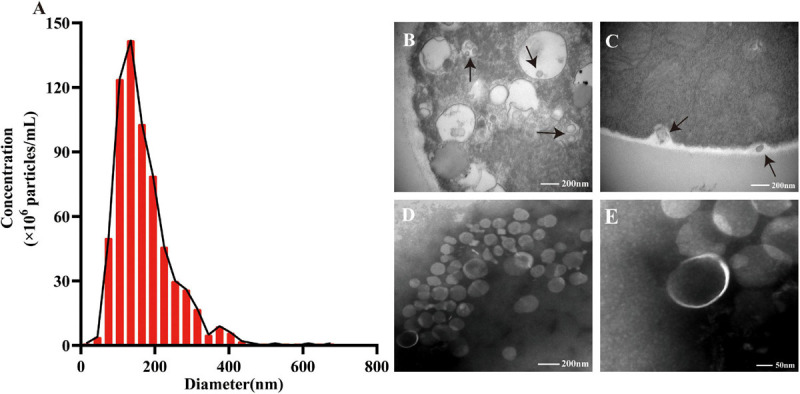
FIGURE 1.

The size distribution and morphology of extracellular vesicles (EVs) secreted by Talaromyces marneffei (TM). (A) Nanoparticle tracking analysis revealed the size distribution and particle concentration of isolated EVs. (B) The EVs were observed inside TM through transmission electron microscopy (TEM; black arrow). (C) It showed the moment of TM yeast cells releasing EVs (black arrow). (B,C) Scale bar = 200 nm. (D,E) TEM identified the purified EVs’ morphology with a cup-shaped structure. (D) Scale bar = 200 nm. (E) Scale bar = 50 nm.
