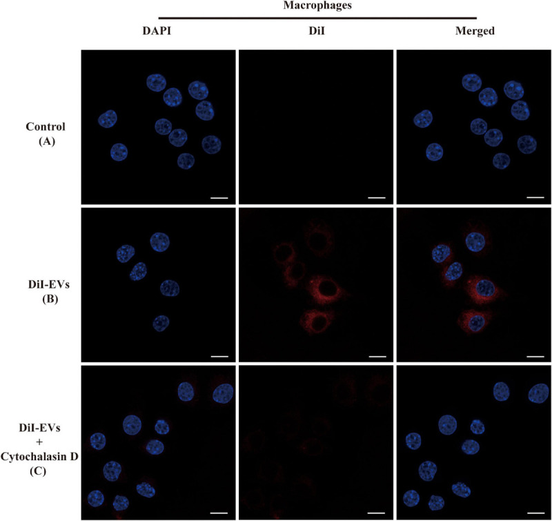
FIGURE 2.

Immunofluorescence images showed the process of RAW264.7 macrophage cells uptaking extracellular vesicles (EVs). The RAW264.7 macrophage cells were incubated with unstained EVs (A) and labeled vesicles (B) after 2 h of co-incubation. (C) RAW264.7 macrophage cells were incubated with actin polymerization inhibitors cytochalasin D for 2 h before the addition of EVs. EVs were labeled with DiI staining (red). Cell nuclei were counterstained with 4′,6-diamidino-2-phenylindole (blue). The fluorescent images are representative of three independent experiments (scale bar = 10 μm).
