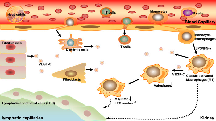
Fig. 9. Schematic diagram of how macrophages participate in lymphangiogenesis in renal fibrosis.
When renal fibrosis is initiated, circulating inflammatory cells are chemically induced to home to the tissue, including macrophages, T cells, dendritic cells, and so on. Macrophages are polarized to M0, M1, and M2 phenotypes. Among them, M1 macrophages can preferentially transdifferentiate into LECs. During this process, the lymphatic growth factor VEGF-C, which is produced by infiltrated inflammatory cells, tubular cells, and other interstitial cells, suppresses M1 macrophage autophagy, thus upregulating M1 marker expression and polarization and ultimately promoting macrophage differentiation into LECs.

