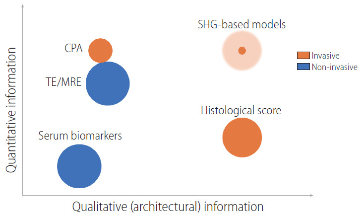
Figure 2.

Comparison of the various noninvasive and invasive methods for fibrosis assessment in terms of the quantitative and qualitative information yielded. The size of the circle represents current utility in clinical practice/trials (shaded area represents potential growth). CPA, collagen proportionate area; TE, transient elastography; MRE, magnetic resonance elastography; SHG, second harmonic generation.
