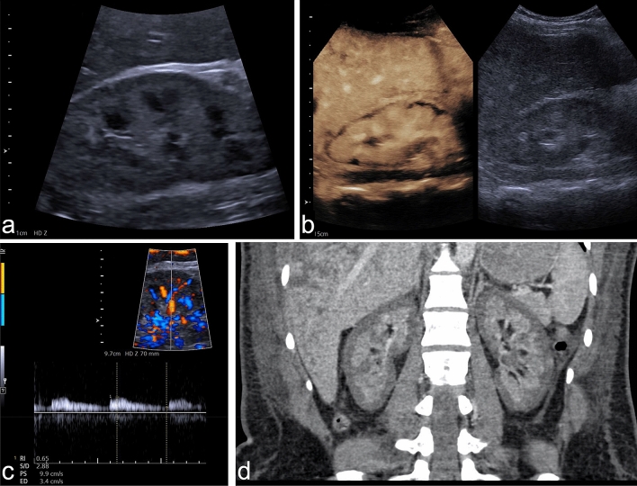
Figure 1.
Example of RCN of the native kidney in a 28-year-old woman suffering from massive postpartum bleeding with acute kidney failure and HELLP syndrome. (a) B-mode image of the right kidney showing a hypoechoic rim of 3–4 mm. (b) CEUS of the right kidney showing a subcapsular loss of contrast enhancement of 3–5 mm. (c) Triplex sonography of the right kidney with a PW spectrum of an interlobar artery showing a normal resistance index of 0.65. (d) Coronal venous-phase CT scan obtained 14 days before CEUS examination showing a recess of contrast agent measuring up to 6 mm in both kidneys, confirming the diagnosis of RCN. CEUS denotes contrast-enhanced ultrasound, RCN, renal cortical necrosis, HELLP syndrome denotes hemolysis, elevated liver enzymes, low platelets, PW, pulsed-wave Doppler.

