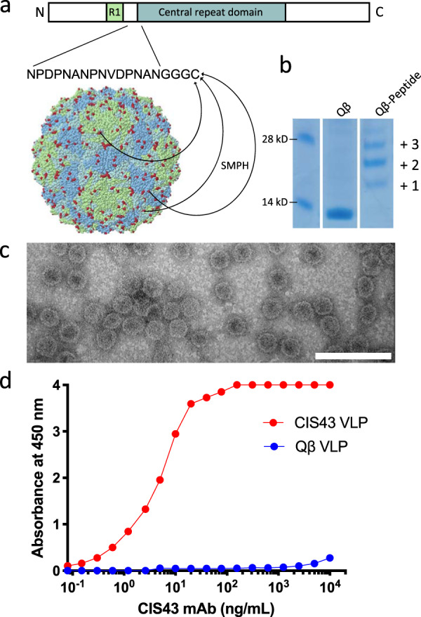
Fig. 1. Characterization of CIS43 VLPs.

a Schematic representation of CSP showing the location of the CIS43 epitope and the process of CIS43 VLP conjugation. A 15-amino acid peptide representing the CIS43 mAb epitope was synthesized to include a (Glycine)3-Cysteine linker and conjugated to surface-exposed lysine residues (shown in red) on the coat protein of Qβ bacteriophage VLPs using the bifunctional crosslinker SMPH. b SDS-PAGE analysis of unconjugated (center lane) or CIS43 peptide conjugated (right lane) Qβ VLPs. The ladder of bands in the CIS43 VLP lane reflect individual copies of coat protein modified with 1, 2, or more copies of the CIS43 peptide. Gel images are derived from the same experiment and were processed in parallel. Size markers are shown in the left lane. The unmodified gel is shown in Supplementary Fig. 2. c Transmission electron micrograph of the CIS43 VLPs. VLPs are visualized at a magnification of 200,000x. Scale bar (in white) represents 100 nm. d Binding of the CIS43 mAb to CIS43 VLPs (red) or wild-type (unmodified) Qβ VLPs (blue) as measured by ELISA.
