
Figure 1.
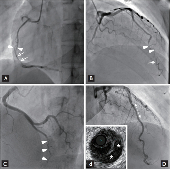
Angiographic classification of spontaneous coronary artery dissection. (A) Patient no. 9: type I, arterial wall contrast staining (arrows) or multiple radiolucent lumens (arrowheads). (B) Patient no. 7: type 2a, abrupt caliber reduction (arrowheads) with restoration of distal vessel caliber (arrow). (C) Patient no. 13: type 2b, abrupt caliber reduction without restoration of distal vessel caliber (arrowheads). (D) Patient no. 5: type 3, tubular stenosis mimicking atherosclerosis (double-point arrow), and confirming false lumen (asterisks) by intravascular ultrasound (d).