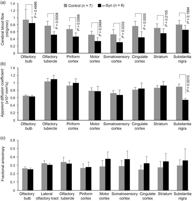
Figure 3.
Plots of quantitative data illustrating changes in CBF (a), ADC (b), and FA (c) in various regions of interest (ROI). All cortical ROI of α-synuclein mice showed significant reductions (P < 0.05) in CBF. [AQ: Please check edit: ‘However, the CBF reduction in the olfactory bulb and the two subcortical ROIs…’]However, the CBF reduction in the olfactory bulb and the two subcortical ROIs (striatum and substantia nigra) was not significant (P > 0.05). None of the ROIs showed a significant change in either ADC or FA, except the substantia nigra, in which the ADC value of α-synuclein mice was significantly reduced (P < 0.05). Data represent mean ± SD. Two-way ANOVA with Tukey-Kramer method of correction for multiple comparisons. The olfactory bulb CBF was excluded from the two-way ANOVA due to being confounded by the use of isoflurane during MRI. Olfactory bulb CBF: unpaired t-test with Welch correction.

