Figure 6.
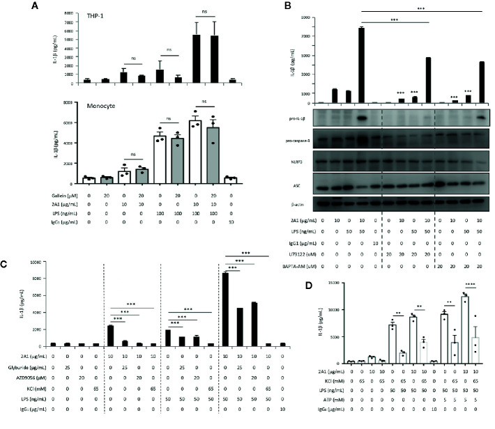
EMR2-mediated signaling in THP-1 cells induces a Gβγ-independent PLC-β activation and Ca2+ mobilization signaling activity, leading to K+ efflux and NLRP3 inflammasome activation. (A, B) Culture supernatants of THP-1 cells (A and B top panel) and monocytes (A, bottom panel) treated with indicated conditions for 24 h were collected for the detection of IL-1β by ELISA and relevant NLRP3 inflammasome proteins by western blotting analyses (B, bottom panel). (C, D) Culture supernatants of THP-1 cells (C) and monocytes (D) treated with indicated conditions for 24 h were collected for the detection of IL-1β by ELISA. Data are means ± SEM of at least three independent experiments performed in triplicate in THP-1 cells (A–C) or monocytes from three different donors (A, D). Data were analyzed by one-way ANOVA. ns, non-significant, ** p<0.01, *** p< 0.001, **** p<0.0001 versus the control group. Gallein, Gβγ inhibitor; U73122, PLC inhibitor; BAPTA-AM, Ca2+ chelator; Glyburide, inhibitor of the sulfonylurea receptor 1 (SUR1) subunit of the ATP-sensitive potassium channels; AZD9056, a selective P2X7 receptor antagonist; KCl, high extracellular KCl concentration served to inhibit cellular K+ efflux.

