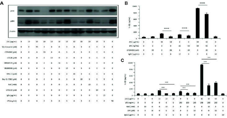
Figure 8.
EMR2-mediated signaling in THP-1 cells induces Akt activation and ROS production, leading to NLRP3 inflammasome activation. (A) Western blotting analyses of EMR2-mediated signaling in THP-1 cells incubated with or without 2A1 and specific signaling inhibitors as indicated for 30 min. Blots were probed to detect phospho-Akt, phospho-ERK and β-actin level. Cells treated with mouse IgG1 and LPS were included as negative and positive controls, respectively. (B, C) Culture supernatants of THP-1 cells treated with indicated conditions for 24 h were collected for the detection of IL-1β by ELISA. Data are means ± SEM of at least three independent experiments performed in triplicate and analyzed by one-way ANOVA. *** p< 0.001, **** p<0.0001 versus the control group. LY294002, PI3K inhibitor; NAC (N-Acetyl-L-cysteine) and DPI (Diphenyleneiodonium), ROS inhibitors.

